page contents

sites

Monday, 28 January 2013

Disease Kidney

Source(google.com.pk)
Disease Kidney Biography
It depends on the type of magnetic resonance imaging (MRI) scan you're scheduled to have — an MRI with contrast or an MRI without contrast. Contrast agents, including gadolinium, are used to enhance some MRI scans. Contrast agents are injected into a vein in your hand or arm. Not all MRIs require a contrast agent.

There are no special concerns for people with kidney problems having an MRI without contrast.

However, there are concerns if people with kidney problems are given a gadolinium-based contrast agent during their MRI — especially if they have severe kidney failure (renal insufficiency).

Gadolinium-containing contrast agents may increase the risk of a rare, but serious, disease called nephrogenic systemic fibrosis in people with severe kidney failure. Nephrogenic systemic fibrosis triggers thickening of the skin, organs and other tissues. There's no effective treatment for this serious, debilitating disease.

Before you have an MRI, ask your doctor if a gadolinium-based contrast agent will be used. If the answer is yes, tell your doctor about your history of kidney problems. If possible, your doctor may select a different imaging test. In cases where an MRI with gadolinium is necessary despite the potential risks, your doctor may use the lowest possible dose of the form of gadolinium that has been associated with the fewest complications as well as consider hemodialysis immediately after the MRI with gadolinium.
Disease Kidney
Disease Kidney
Disease Kidney
Disease Kidney

Disease Kidney
Disease Kidney
Disease Kidney
Disease Kidney
Disease Kidney
Disease Kidney

Artificial Kidney

Source(google.com.pk)
Artificial Kidney Biography
An additional line of research on renal regeneration focuses on development of living membranes for an intradialytic biological kidney support device. End-stage renal disease patients have uremic complications that result in high cardiovascular morbidity and a poor quality of life, despite hemodialysis. Uremia is caused by the retention of a large group of molecules with different physical and chemical properties that are not sufficiently cleared by hemodialysis. Within REFORM, a cell device (BioKid) will be developed capable of effective clearance of these toxins ex vivo. The BioKid will comprise of multiple so-called living membranes, i.e. tight monolayers of human renal epithelial cells that are grown on newly designed semi-permeable bioactive polymer membranes. A unique supramolecular approach will be used to develop a 2D bioactive polymer membrane that regulates long-lived monolayer integrity and cell viability under uremic conditions. The expertise and knowledge gained on the supramolecular 2D bioactive polymer membrane will be translated into a 3D configuration that will be applied in a simple in vitro set-up as a cell-aided intradialytic uremic toxin removal device. This will serve as a proof of principle for a more sophisticated device that can be used in the future to treat uremic symptoms in end-stage renal disease patients on dialysis.

Chronic Renal Failure (CRF)/Kidney Disease

Source(google.com.pk)
Chronic Renal Failure (CRF)/Kidney Disease Biography

Chronic Renal Failure (CRF) or kidney disease is so prevalent in older cats today, this case study is so important from a holistic perspective. Here is Kent's story:

 It was nearly 17 years ago when I brought home 3 adorable Himalayan kittens: Rocky, Kasey and Chanda. Rocky and Kasey littermates and Chanda had the same father. For their entire lives I gave them the best care I knew of — following the advice of a respected, well-established veterinarian clinic, I gave them the highest quality food touted on the market like Iams and Hills Science Diet and lots of TLC!

This past summer I learned the other side of pet ownership. In August 2006, we learned Rocky had intestinal cancer and within 4 weeks he was gone. One week later, I took Kasey in for a senior screening and learned he was in the advanced stages of Chronic Renal Failure (CRF). [Sigh] Kasey did great, however, and for 4 months we enjoyed some of the best times of his life! He really was mostly “normal” clinically right up until the day before his death on Dec 25th. Amazing!

Several years ago I had done some extensive study as a non-practitioner in the area of human nutrition, supplements, antioxidants and free radicals and their role in various diseases, cancer, and the aging process. I had thought because the cat food bag said “Antioxidants” on the outside I was giving them proper nutrition. I had much to learn. I began searching the Internet for information related to cancer and CRF in cats and found myself once again learning volumes on the role that nutrition, lifestyle and antioxidants play in life—only this time it was related to cats. I started with Feline CRF Information Center which is absolutely fantastic and filled with so much practical and helpful information! On their site I discovered LittleBigCat.com and the excellent work of Dr. Jean Hofve, DVM, who has turned her practice to the extensive study of nutrition and lifestyle in cats. EVERY CAT OWNER NEEDS TO STUDY THE INFORMATION ON HER WEBSITE!

Among the wealth of information I gleaned from Little Big Cat, two areas were paramount. The importance of canned or wet food over dry food and bio-algae concentrates for whole food supplementation. I encourage you to read through the information on the above websites but here is the short version of these two important points:

1. The importance of canned or wet food (or even raw) over dry food—cats are by nature arid (dry climate) animals and thus are designed by their Creator to obtain the majority of their moisture from the foods they eat (i.e. birds, mice, rats, etc.). Do the math—give your cats dry food and you are compounding the dehydration factor which can lead to numerous health issues including but not limited to CRF.

2. Bio-algae concentrates supplementation—I don’t sell the stuff so you can put your guard down right away as this is not an attempt to sell you anything. From what I have learned, cats, being obligate carnivores, do not have the ability to break down the cell walls of plants thus it is probably a waste of time to give them fruits & veggies. However, the bio-algae concentrates are unique in that cats are in fact able to process it and obtain an incredible pallet of antioxidants, essential fatty acids (Omega 3, 6, 9 and GLA oils), over 4,000 enzymes, vitamins, amino acids, all known minerals and trace elements, etc.

Quickly then to my remaining 16+ yr old cat, Chanda—just give her the canned food with added bio-algae concentrates right? Wrong! As you know, cats are creatures of habit and very finicky. They will not just switch foods or accept something new thrown in. Dr. Jean Hofve has written an article suggesting how to go about switching food as well as one on bio-algae concentrates I encourage you to read both.

What worked for Chanda: I continued to supply her with the dry food she had been eating, and started putting the canned food out, Life's Abundance’s Instinctive Choice formulated by holistic veterinarian, Dr. Jane Bicks. I carefully followed the suggestions of Russell Louie of Optimum Choices, LLC where he suggested starting bio-algae concentrates with only the amount that will fit on the end of a flat toothpick and gradually increasing another toothpick measure every 3-5 days or so. It took five months of patience and persistence, throwing a lot of food away, but I had made the commitment up front, realizing that 83 cents per can of food plus an initial bottle of bio-algae concentrates was a small price to pay even if I had to throw much of it away during the first few months because Chanda’s health and vitality is worth it! If it needs to be a fresh can each meal, who cares? Again, 83 cents a can is a small price to pay compared to the cost of veterinary care. I used glass Pyrex storage containers for their food and water bowls as well as food storage of the canned food. In doing so, you avoid the risk of leaching contaminants from plastic or metal containers into the food and supposedly the food tastes better to cats.

Chanda today: despite having been diagnosed with CRF, her numbers/kidney values are stable, her blood pressure is normal, eyes are clear, and clinically she literally has as much energy and spirit as her earlier years, once again playing with her toys and running through the house. She has such a soft and shiny coat that everyone that meets her, whether a veterinarian, vet tech, stranger or friend remarks about her soft coat! They simply cannot believe she is nearly 19 yrs old (2009)!

Please realize I am not suggesting that switching to canned food or adding bio-algae concentrates are “miracle” cures and I am not a veterinarian. I am simply letting you know what my experience has been, in the hopes you will get the same or similar results with your pet! I know Chanda will not be with me forever—I may have weeks, months, a year or at most several years given the average life of a cat is 15-16 years but I want however much time she has to be as enjoyable and healthy as possible. Whether the above is adding years to her life, I do not know but I do know it is adding life to her years!

Kidney Diseases

Source(google.com.pk)
Kidney Diseases Biography

After our first two cats died of kidney disease or Chronic Renal Failure (CRF) at age 17, we learned that grains are not an appropriate diet for carnivores (meat-eating animals needing 60-90% meat). Chronic Renal Failure (CRF) is almost an epidemic among older cats because so many people feed dry kibble in free-feeders to their felines (unnatural for wild cats). Some holistic veterinarians say kidney disease could almost be prevented if cats were not fed dry kibble all their lives. Dry kibble must contain grains or other carbohydrates to hold it together during processing. At most, premium brands contain only 50% meat (some new brands now claim 80%) and most (cheaper) brands are less than 30%. Upon learning that, we transitioned our remaining cat to a grain-free canned and raw food diet.

When we acquired our dog (needs 60-80% meat) we immediately put her on a canned food diet and then transitioned her to raw food. Even the training treats we give our dog are grain-free (we buy dried meat treats sold at natural pet stores instead of dog “cookies”).

One reason to avoid grains is the quality in most commercial pet foods is different than what you would cook for your pet. Typically, the good quality grains go into human foods and what is left over (unfit for human consumption) gets used in pet food. The grains in pet food are more likely to contain toxic molds, pesticides and man-made contaminants (such as melamine). Also, the plant protein in grains and soy in pet food gets counted towards the total protein of the food, though our pets, as carnivores, can’t utilize it. They are designed to get their protein from meat, not plants. The holistic veterinarian and pet food expert Dr. Jean Hofve recommends avoiding all non-meat protein sources. She states that cheap vegetable protein substitutes are inappropriate in the diet of a carnivore and are used by the pet food companies only to increase profit.

From our holistic perspectives, based on our research and according to many other holistic veterinarians, we do not subscribe to the low-protein diet for chronic renal failure cats. This low-protein diet myth was originally based on erroneous studies in the 1920-30's. The benefits of low-protein diets were never scientifically proven but often accepted. Click this link to read what a vet has to say, Mythology of Protein Restriction for Dogs with Reduced Renal Function. Most holistic vets now suggest feeding a high quality protein diet to chronic renal failure cats. As CRF cats get pickier in their tastes, it is more important to get good premium quality meat protein into their bodies, than worry about any percentages.

It is especially important to avoid grains with cats because they are “obligate carnivores” (must have meat to survive). Their bodies are not designed to digest carbohydrates. Cats need 70-90% meat (protein + fat) and cannot survive as vegetarians. Domestic cats are descended from the African wild cat, whose natural diet was limited to rodents, birds, eggs, reptiles and insects. This wild cat was domesticated by the Egyptians 4,000 years ago to protect their granaries from rodents. The cat was ideally suited to this task, since it did not eat grains. They got their moisture from their food and not from drinking water, as they lived in the desert. Their prey contains 65-75 percent water. Cats do not have a strong thirst drive compared to other mammals so they will not drink water until they are already dehydrated.

Yet today, most people feed their cats dry kibble that contain more grains than meat and only averages 10 percent water. Could this be why so many cats get kidney disease or Chronic Renal Failure (CRF)? The new grain-free kibbles are not a solution either since they lack water and the higher protein in them makes them even more dehydrating. They still have a fattening starch in them such as potato or tapioca starch to hold them together. The epidemic of obesity in our dogs and cats, which can cause many health problems, is yet another reason not to feed our pets grains or other starches (which are fattening to carnivores). Other health problems caused by feeding dry kibble include urinary tract disorders and diabetes. One holistic vet called dry kibble for cats “diabetes in a bag” and holistic vets state that diabetes can often be cured by taking the cat off of dry food.

To learn more about the optimum diet for cats (and dogs), see our e-Book and the CD audio seminar How NOT to Kill Your Cat or Dog by Dr. Jean Hofve. Dr. Jean Hofve recommends supplements high in Omega-3 fatty acids and antioxidants for cats with kidney disease. Not only does bio-algae concentrates  have Omega-3 fatty acids but it also contains Omega-6, Omega-9, GLAs (Gamma-Linolenic Acids), ALAs (Alpha-Linolenic Acids), DGLA (Dihomogamma-Linolenic Acid), DHA (Docosahexaenoic acid) and more fatty acids. Just as important as these individual fatty acid ingredients, is the fact that they are all in the proper proportions and dosages that Nature intended animals to get. This is the synergy found in whole food products not present in isolated, extracted and man-made fatty acids supplements. One can literally imbalance the body further by treating the symptoms with just an Omega-3 fatty acid supplement.

As far as antioxidants, bio-algae concentrates (Spirulina pacifica, Spirulina platensis, Dunaliella salina and astaxanthin from Haematococcus pluvialis) contain some of the world's most powerful: beta-carotene, alpha-carotene, gamma-carotene, lycopene, lutein, beta-cryptoxanthin, zeaxanthin, and astaxanthin. Astaxanthin has 500-1000 times the antioxidant capacity of Vitamin E, greater anti-inflammatory capability than Vitamin E, 40 times the antioxidant capacity of beta-carotene and almost 4 times the antioxidant capacity of lutein. The antioxidants in bio-algae concentrates are so good, we saw a mast cell cancer tumor on a dog disappear in ten weeks using only 1.5 capsules/day.

Here are some case studies from customers whose cats had CRF (Chronic Renal Failure) and used bio-algae concentrates (Spirulina pacifica, Spirulina platensis, Dunaliella salina and astaxanthin from Haematococcus pluvialis).
Kidney Diseases 

Kidney Diseases 

Kidney Diseases 

Kidney Diseases 

Kidney Diseases 
Kidney Diseases 

Kidney Diseases 

Kidney Diseases 

Kidney Diseases 

Kidney Diseases 

Kidney Diseases 

Renal Kidney

Source(google.com.pk)
Renal Kidney Biography

End-stage renal disease, or chronic kidney failure, affects more than 500,000 people per year in the United States alone and is only fully treated with a kidney transplant.

Yet there were only 17,000 donated kidneys for transplants last year and the number of patients on the transplant waiting list currently exceeds 85,000, according to the Organ Procurement and Transplant Network.

350,000 patients are instead reliant on kidney dialysis, which comes at a tremendous cost. The Medicare system alone spends $25 billion on treatments for kidney failure, more than 6 percent of the total budget, though the disease affects only 1 percent of Medicare recipients. That cost includes almost $75,000 per patient each year for dialysis, according to the U.S. Renal Data System.   Dialysis also takes a human toll. A typical dialysis schedule is three sessions per week, for 3 to 5 hours per session, in which blood is pumped through an external circuit for filtration. This is exhausting for patients and only replaces 13 percent of kidney function and, as a result, only 35 percent of patients survive for more than 5 years.

With the limited supply of donors, that means thousands of patients die each year waiting for a kidney, but UCSF researchers today unveiled a prototype model of the first implantable artificial kidney, in a development that one day could eliminate the need for dialysis.

The device, which would include thousands of microscopic filters as well as a bioreactor to mimic the metabolic and water-balancing roles of a real kidney, is being developed in a collaborative effort by engineers, biologists and physicians nationwide, led by Shuvo Roy, PhD, in the UCSF Department of Bioengineering and Therapeutic Sciences.

The treatment has been proven to work for the sickest patients using a room-sized external model developed by a team member in Michigan. Roy's goal is to apply silicon fabrication technology, along with specially engineered compartments for live kidney cells, to shrink that large-scale technology into a device the size of a coffee cup. The device would then be implanted in the body without the need for immune suppressant medications, allowing the patient to live a more normal life.


A model of the implantable bioartificial kidney shows the two-stage system. Thousands of nanoscale filters remove toxins from the blood, while a BioCartridge of renal tubule cells mimics the metabolic and water-balance roles of the human kidney.  Credit: UCSF

"This device is designed to deliver most of the health benefits of a kidney transplant, while addressing the limited number of kidney donors each year," said Roy, an associate professor in the UCSF School of Pharmacy who specializes in developing micro-electromechanical systems (MEMS) technology for biomedical applications. "This could dramatically reduce the burden of renal failure for millions of people worldwide, while also reducing one of the largest costs in U.S. healthcare."

The team has established the feasibility of an implantable model in animal models and plans to be ready for clinical trials in five to seven years.

The two-stage system uses a hemofilter to remove toxins from the blood, while applying recent advances in tissue engineering to grow renal tubule cells to provide other biological functions of a healthy kidney. The process relies on the body's blood pressure to perform filtration without needing pumps or an electrical power supply.

The project exemplifies the many efforts under way at UCSF to build collaborations across scientific disciplines that accelerate the translation of academic research into real solutions for patients, according to Mary Anne Koda-Kimble, PharmD, dean of the UCSF School of Pharmacy.

"This is a perfect example of the work we are doing at UCSF to address some of the most critical medical issues of our time, both in human and financial costs," Koda-Kimble said. "This project shows what can be accomplished by teams of scientists with diverse expertise, collaborating to profoundly and more quickly improve the lives of patients worldwide."

The creation of the Department of Bioengineering and Therapeutic Sciences – a joint department in the UCSF schools of Pharmacy and Medicine – was itself an effort to promote translational research at UCSF by forming collaborations across biomedical specialties. Roy is also a founding faculty member of the UCSF Pediatric Device Consortium, which aims to accelerate the development of innovative devices for children health, and a faculty affiliate of the California Institute for Quantitative Biosciences (QB3) at UCSF.

His team is collaborating with 10 other teams of researchers on the project, including the Cleveland Clinic where Roy initially developed the idea, Case Western Reserve University, University of Michigan, Ohio State University, and Penn State University.

The first phase of the project, which has already been completed, focused on developing the technologies required to reduce the device to a size that could fit into the body and testing the individual components in animal models. In the second and current phase, the team is doing the sophisticated work needed to scale up the device for humans. The team now has the components and a visual model and is pursuing federal and private support to bring the project to clinical use.
Renal Kidney

Renal Kidney

Renal Kidney

Renal Kidney

Renal Kidney

Renal Kidney

Renal Kidney

Renal Kidney

Renal Kidney

Renal Kidney

Renal Kidney

Wednesday, 23 January 2013

Renal And Kidney

Source(google.com.pk)
Renal And Kidney Biography

The principal function of the urinary system is to maintain the volume and composition of body fluids within normal limits. One aspect of this function is to rid the body of waste products that accumulate as a result of cellular metabolism. Other aspects of its function include regulating the concentrations of various electrolytes in the body fluids and maintaining normal pH of the blood.

In addition to maintaining fluid homeostasis in the body, the urinary system controls red blood cell production by secreting the hormone erythropoietin. The urinary system also plays a role in maintaining normal blood pressure by secreting the enzyme renin.

The urinary system consists of the kidneys, ureters, urinary bladder, and urethra. The kidneys form the urine and account for the other functions attributed to the urinary system. The ureters carry the urine away from kidneys to the urinary bladder, which is a temporary reservoir for the urine. The urethra is a tubular structure that carries the urine from the urinary bladder to the outside.

Kidneys
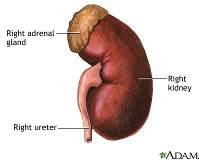
The kidneys are the primary organs of the urinary system. The kidneys are the organs that filter the blood, remove the wastes, and excrete the wastes in the urine. They are the organs that perform the functions of the urinary system. The other components are accessory structures to eliminate the urine from the body.

The paired kidneys are located between the twelfth thoracic and third lumbar vertebrae, one on each side of the vertebral column. The right kidney usually is slightly lower than the left because the liver displaces it downward. The kidneys protected by the lower ribs, lie in shallow depressions against the posterior abdominal wall and behind the parietal peritoneum. This means they are retroperitoneal. Each kidney is held in place by connective tissue, called renal fascia, and is surrounded by a thick layer of adipose tissue, called perirenal fat, which helps to protect it. A tough, fibrous, connective tissue renal capsule closely envelopes each kidney and provides support for the soft tissue that is inside.


In the adult, each kidney is approximately 3 cm thick, 6 cm wide, and 12 cm long. It is roughly bean-shaped with an indentation, called the hilum, on the medial side. The hilum leads to a large cavity, called the renal sinus, within the kidney. The ureter and renal vein leave the kidney, and the renal artery enters the kidney at the hilum.

The outer, reddish region, next to the capsule, is the renal cortex. This surrounds a darker reddish-brown region called the renal medulla. The renal medulla consists of a series of renal pyramids, which appear striated because they contain straight tubular structures and blood vessels. The wide bases of the pyramids are adjacent to the cortex and the pointed ends, called renal papillae, are directed toward the center of the kidney. Portions of the renal cortex extend into the spaces between adjacent pyramids to form renal columns. The cortex and medulla make up the parenchyma, or functional tissue, of the kidney.

The central region of the kidney contains the renal pelvis, which is located in the renal sinus and is continuous with the ureter. The renal pelvis is a large cavity that collects the urine as it is produced. The periphery of the renal pelvis is interrupted by cuplike projections called calyces. A minor calyx surrounds the renal papillae of each pyramid and collects urine from that pyramid. Several minor calyces converge to form a major calyx. From the major calyces the urine flows into the renal pelvis and from there into the ureter.

Each kidney contains over a million functional units, called nephrons, in the parenchyma (cortex and medulla). A nephron has two parts: a renal corpuscle and a renal tubule. The renal corpuscle consists of a cluster of capillaries, called the glomerulus, surrounded by a double-layered epithelial cup, called the glomerular capsule. An afferent arteriole leads into the renal corpuscle and an efferent arteriole leaves the renal corpuscle. Urine passes from the nephrons into collecting ducts then into the minor calyces.

The juxtaglomerular apparatus, which monitors blood pressure and secretes renin, is formed from modified cells in the afferent arteriole and the ascending limb of the nephron loop.

Ureter
Each ureter is a small tube, about 25 cm long, that carries urine from the renal pelvis to the urinary bladder. It descends from the renal pelvis, along the posterior abdominal wall, behind the parietal peritoneum, and enters the urinary bladder on the posterior inferior surface.

The wall of the ureter consists of three layers. The outer layer, the fibrous coat, is a supporting layer of fibrous connective tissue. The middle layer, the muscular coat, consists of inner circular and outer longitudinal smooth muscle. The main function of this layer is peristalsis to propel the urine. The inner layer, the mucosa, is transitional epithelium that is continuous with the lining of the renal pelvis and the urinary bladder. This layer secretes mucus which coats and protects the surface of the cells.

Urinary Bladder
The urinary bladder is a temporary storage reservoir for urine. It is located in the pelvic cavity, posterior to the symphysis pubis, and below the parietal peritoneum. The size and shape of the urinary bladder varies with the amount of urine it contains and with pressure it receives from surrounding organs.

The inner lining of the urinary bladder is a mucous membrane of transitional epithelium that is continuous with that in the ureters. When the bladder is empty, the mucosa has numerous folds called rugae. The rugae and transitional epithelium allow the bladder to expand as it fills.

The second layer in the walls is the submucosa that supports the mucous membrane. It is composed of connective tissue with elastic fibers.

The next layer is the muscularis, which is composed of smooth muscle. The smooth muscle fibers are interwoven in all directions and collectively these are called the detrusor muscle. Contraction of this muscle expels urine from the bladder. On the superior surface, the outer layer of the bladder wall is parietal peritoneum. In all other regions, the outer layer is fibrous connective tissue.


There is a triangular area, called the trigone, formed by three openings in the floor of the urinary bladder. Two of the openings are from the ureters and form the base of the trigone. Small flaps of mucosa cover these openings and act as valves that allow urine to enter the bladder but prevent it from backing up from the bladder into the ureters. The third opening, at the apex of the trigone, is the opening into the urethra. A band of the detrusor muscle encircles this opening to form the internal urethral sphincter.

Urethra
The final passageway for the flow of urine is the urethra, a thin-walled tube that conveys urine from the floor of the urinary bladder to the outside. The opening to the outside is the external urethral orifice. The mucosal lining of the urethra is transitional epithelium. The wall also contains smooth muscle fibers and is supported by connective tissue.

The internal urethral sphincter surrounds the beginning of the urethra, where it leaves the urinary bladder. This sphincter is smooth (involuntary) muscle. Another sphincter, the external urethral sphincter, is skeletal (voluntary) muscle and encircles the urethra where it goes through the pelvic floor. These two sphincters control the flow of urine through the urethra.

In females, the urethra is short, only 3 to 4 cm (about 1.5 inches) long. The external urethral orifice opens to the outside just anterior to the opening for the vagina.

In males, the urethra is much longer, about 20 cm (7 to 8 inches) in length, and transports both urine and semen. The first part, next to the urinary bladder, passes through the prostate gland and is called the prostatic urethra. The second part, a short region that penetrates the pelvic floor and enters the penis, is called the membranous urethra. The third part, the spongy urethra, is the longest region. This portion of the urethra extends the entire length of the penis, and the external urethral orifice opens to the outside at the tip of the penis.

Renal And Kidney
Renal And Kidney
Renal And Kidney
Renal And Kidney
Renal And Kidney
Renal And Kidney
Renal And Kidney
Renal And Kidney
Renal And Kidney
Renal And Kidney
Renal And Kidney

Kidney

Source(google.com.pk)
Kidney Biography
Chronic kidney disease is a general term for heterogeneous disorders affecting kidney structure and function. The 2002 guidelines for definition and classification of this disease represented an important shift towards its recognition as a worldwide public health problem that should be managed in its early stages by general internists. Disease and management are classified according to stages of disease severity, which are assessed from glomerular filtration rate (GFR) and albuminuria, and clinical diagnosis (cause and pathology). Chronic kidney disease can be detected with routine laboratory tests, and some treatments can prevent development and slow disease progression, reduce complications of decreased GFR and risk of cardiovascular disease, and improve survival and quality of life. In this Seminar we discuss disease burden, recommendations for assessment and management, and future challenges. We emphasise clinical practice guidelines, clinical trials, and areas of uncertainty.

Screening (looking) for early kidney disease in people who are not already known to have it. Kidney disease is common and is commonly insidious in onset. The burden of kidney disease in its earlier stages lies not only in the risk of progression but in the complications of decreased kidney function and the risk of heart disease.

In 2002 the National Kidney Foundation (NKF) set forth guidelines for kidney disease screening. It recommended that all individuals at increased risk for chronic kidney disease have their blood pressure measured and their blood and urine tested for signs of impaired kidney function. Those at increased risk for chronic kidney disease were defined as people with:

Kidney
Kidney
Kidney
Kidney
Kidney
Kidney
Kidney
Kidney
Kidney
Kidney
Kidney
Kidney