page contents

sites

Wednesday 2 October 2013

Tips From Former Smokers

Tips From Former Smokers Biography

Source(google.com.pk)
The individuals below are participating in the Tips From Former Smokers Campaign. All of them have been affected by cigarette smoke. Some are former smokers and some have never smoked. Almost all of them are living with smoking-related diseases and disabilities. These diseases and disabilities changed the quality of their lives — some dramatically — including how they eat, dress and do daily tasks. Some had to give up activities they once loved to do. They speak from experience and agreed to share their stories with you, to send a single, powerful message: Quit smoking now. Or better yet — don't ever start.

Meet Annette Meet Annette. Annette, age 57, lives in New York and began smoking in her teens. At age 52, she was diagnosed with lung cancer, which required removal of one of her lungs. She was later diagnosed with oral cancer.


Meet Beatrice Meet Beatrice. Beatrice, age 40, lives in New York and began smoking regularly at age 13. A mother of two, she quit smoking in 2010 because she wanted to be around for her family.


Meet Bill Meet Bill. Bill, age 40, lives in Michigan and has diabetes. At 15, he started smoking cigarettes. At 39, he quit smoking after his leg was amputated due to poor circulation—made worse from smoking.


Meet Brandon Meet Brandon. Brandon, age 31, lives in North Dakota and began smoking at age 15. At 18, he was diagnosed with Buerger's disease, which resulted in the amputation of both his legs and several fingertips.


Meet Christine Meet Christine. Christine, age 49, lives in Pennsylvania and began smoking at age 16. At age 44, she was diagnosed with oral cancer, which eventually required doctors to remove half of her jaw.


Meet Ellie Meet Ellie. Ellie, age 57, lives in Florida and never smoked. At 35, she started having asthma attacks triggered from breathing secondhand smoke at work. The severe attacks forced her to leave a job she loved.


Meet Jamason Meet Jamason. Jamason, age 18, lives in Kentucky. He was an infant when he was diagnosed with asthma. When people smoke around him, the secondhand smoke can trigger life-threatening asthma attacks.


Meet James Meet James. James, age 48, lives in New York and began smoking at age 14. He quit smoking in 2010 to reduce his risk for health problems and now bikes 10 miles every day.


Meet Jessica Meet Jessica. Jessica, age 28, lives in New York and has never smoked. Her son, Aden, was diagnosed with asthma at age 3, and exposure to secondhand smoke has triggered asthma attacks.


Meet Mariano Meet Mariano. Mariano, 55, lives in Illinois. He started smoking at 15. In 2004, he had open heart surgery and barely escaped having a heart attack. He quit smoking—grateful for a second chance at life.


Meet Marie Meet Marie. Marie, age 62, lives in New York and began smoking in high school. Diagnosed with Buerger's disease in her forties, Marie has undergone amputations of part of her right foot, her left leg, and several fingertips.


Meet Michael Meet Michael. Michael, age 57, lives in Alaska and began smoking at age 9. At 44, he was diagnosed with COPD—chronic obstructive pulmonary disease—which makes it harder and harder to breathe and can cause death.


Meet Nathan Meet Nathan. Nathan, 54, lives in Idaho. A member of the Oglala Sioux tribe, he was exposed to secondhand smoke at work that caused permanent lung damage and triggered asthma attacks so severe he had to leave his job.


Meet Roosevelt Meet Roosevelt. Roosevelt, age 51, lives in Virginia and began smoking in his teens. At age 45, he had a heart attack. Doctors later placed stents in his heart and performed six bypasses.


Meet Shane Meet Shane. Shane lives in Wisconsin and began smoking at age 18. At age 34, he was diagnosed with throat cancer and his larynx and upper esophagus were removed. Today, at age 44, he continues to battle cancer.


Meet Sharon Meet Sharon. Sharon, age 52, lives in Illinois and began smoking at age 13. In 1997, at age 37, she was diagnosed with stage IV throat cancer.


Meet Shawn Meet Shawn. Shawn, age 51, lives in Washington State and began smoking at age 14. At 46, he was diagnosed with throat cancer that required 38 radiation treatments and the removal of his larynx.


Meet Suzy Meet Suzy. Suzy, age 62, lives in New York and began smoking at age 15. At age 57, Suzy suffered a stroke that caused her to have partial paralysis and problems with her speech and eyes.


Meet Terrie Meet Terrie. Terrie lived in North Carolina and began smoking in high school. At 40, she was diagnosed with oral and throat cancers and had her larynx removed. She died of a smoking-related cancer on September 16, 2013. She was 53.


Meet Tiffany Meet Tiffany. Tiffany, age 35, lives in Louisiana. She started smoking at 19, even though her mother, a smoker, died of lung cancer. Tiffany quit smoking—wanting to be around for her own teenage daughter.


Meet Wilma Meet Wilma. Wilma, age 49, lives in Texas and began smoking in her early teens. She quit smoking in 2007 to reduce her risk for health problems.

Mouth Cancer Symptoms From Smoking

Mouth Cancer Symptoms From Smoking Biography

Source(google.com.pk)
Mouth Cancer Symptoms From Smoking

Mouth Cancer Symptoms From Smoking


What Is Cancer?
Cancer refers to diseases in which abnormal cells divide out of control and are able to invade other tissues. Cancer cells can spread to other parts of the body through the blood and lymph systems.1,2
There are more than 100 different types of cancer. Most cancers are named for the organ or type of cell in which they start — for example, lung cancer begins in the lung and laryngeal cancer begins in the larynx (voice box).1
Symptoms can include:3
A thickening or lump in any part of the body
Weight loss or gain with no known reason
Sores that won't heal
A new mole or a changes in an existing mole
Hoarseness or a cough that does not go away
A hard time swallowing
Indigestion or pain after eating that does not go away
Changes in bowel or bladder habits
Unusual bleeding or discharge
Feeling weak or very tired

How Is Smoking Related to Cancer?
Smoking can cause cancer and then block your body from fighting it:4
Poisons in cigarette smoke can weaken the body’s immune system, making it harder to kill cancer cells. When this happens, cancer cells keep growing without being stopped.
Poisons in tobacco smoke can damage or change a cell"s DNA. DNA is the cell"s "instruction manual" that controls a cell"s normal growth and function. When DNA is damaged, a cell can begin growing out of control and create a cancer tumor.
Doctors have known for years that smoking causes most lung cancer. Nearly 9 out of 10 men who die from lung cancer are smokers. And about 3,000 nonsmokers die each year from lung cancer caused by secondhand smoke.4
Smoking can cause cancer almost anywhere in your body, including the:4
Mouth, nose, and throat
Larynx
Trachea
Esophagus
Lungs
Stomach
Pancreas
Kidneys and ureters
Bladder
Cervix
Bone marrow and blood
Smokeless tobacco also causes cancer, including cancers of the:5
Esophagus
Mouth and throat
Pancreas

How Can Smoking-Related Cancers Be Prevented?
Quitting smoking lowers the risks for cancers of the lung, mouth, throat, esophagus, and larynx.4,6
Within 5 years of quitting, your chance of cancer of the mouth, throat, esophagus, and bladder is cut in half.4
Ten years after you quit smoking, your risk of dying from lung cancer drops by half.4
If nobody smoked, one of every three cancer deaths in the United States would not happen.4

How Is Cancer Treated?
The treatment for cancer depends on the type of cancer and the stage of the disease (how severe the cancer is and whether it has spread). Doctors may also consider the patient's age and general health. Often, the goal of treatment is to cure the cancer. In other cases, the goal is to control the disease or to reduce symptoms for as long as possible. The treatment plan for a person may change over time.

Most treatment plans include surgery, radiation therapy, or chemotherapy. Some plans involve hormone therapy (a treatment to keep cancer cells from getting the hormones they need to grow). Other plans involve biological therapy (a treatment that helps your immune system fight cancer).
Some cancers respond best to a single type of treatment. Other cancers may respond best to a combination of treatments.

For patients who get very high doses of chemotherapy or radiation therapy, a stem cell transplant, also known as a bone marrow transplant, may be recommended by their doctor. This is because high-dose therapies destroy both cancer cells and normal blood cells. A stem cell transplant can help the body to make healthy blood cells to replace the ones lost due to the cancer treatment. It’s a complicated procedure with many side effects and risks.

Mouth cancer Treatment

Mouth cancer Treatment Biography

Source(google.com.pk)
This factsheet is for people who have mouth cancer, or who would like information about it, including symptoms, causes and treatments.

Mouth cancer (oral cancer) is caused by an abnormal and uncontrolled growth of cells in the mouth.

Animation: How cancer develops
About mouth cancer
Types of mouth cancer
Symptoms of mouth cancer
Causes of mouth cancer
Diagnosis of mouth cancer
Treatment of mouth cancer
Prevention of mouth cancer


Animation: How Cancer Develops

About mouth cancer

Mouth cancer includes cancer that starts anywhere in your mouth, including:

your lips
your tongue
your gums
in the floor of your mouth or under your tongue
inside your cheeks and lips
in the roof of your mouth (the palate)
in the area behind your wisdom teeth
Nearly 5,400 people are diagnosed with mouth cancer every year in the UK. Most of these people are over the age of 50, and it affects more men than women.

Types of mouth cancer
Nine out of 10 mouth cancers are squamous cell carcinomas. They develop in the flat, skin-like cells that cover the inside of your mouth. Other, rarer types of mouth cancer include:

salivary gland cancer, which starts in your salivary gland cells
lymphoma, which starts in lymph tissue near the base of your tongue and tonsils
melanoma, which starts in skin pigment cells around your mouth or on your lips
Symptoms of mouth cancer

The two most common symptoms of mouth cancer are:

an ulcer in your mouth or on your lip that won't heal
constant discomfort or pain in your mouth
Other symptoms can include:

red or white patches in your mouth
a lump on your lip, tongue or in your neck
bad breath
unexplained bleeding in your mouth
numbness in your mouth
loose teeth
problems chewing or swallowing, difficulty moving your jaw or a feeling that something is caught in your throat
changes to your voice – it may sound husky or quieter or you may slur your words
weight loss because of problems swallowing
These symptoms aren't always caused by mouth cancer but if you have any of them, see your GP or dentist.

Causes of mouth cancer

The exact reasons why you may develop mouth cancer aren't fully understood at present. However, you may be more likely to develop mouth cancer if you:

smoke any form of tobacco – cigarettes, cigars and pipes, as well as bidis or hand-rolled cigarettes that contain cannabis
chew tobacco, such as betel quid, gutkha and paan
drink excessive alcohol, especially at the same time as smoking or chewing tobacco
have already had certain types of cancer, such as skin cancer
regularly expose yourself to the sun or ultraviolet (UV) light as this increases the risk of lip cancer
have a weakened immune system – people who have HIV/AIDS, or who are taking medicines that suppress the immune system, are more likely to develop mouth cancer
eat a poor diet that doesn’t include many vitamins and minerals
have the human papilloma virus (HPV) as this causes some cancers of the oropharynx (part of the throat at the back of your mouth), including the soft palate, the base of your tongue, and your tonsils

Diagnosis of mouth cancer
The earlier mouth cancer is diagnosed, the better your chances of recovery. Your dentist may spot mouth cancer in its early stages during a routine check-up, so it's important to visit your dentist regularly.

Your GP or dentist will ask about your symptoms and examine you. He or she may also ask you about your medical history. Your GP or dentist will feel your neck and face to check for swellings and may refer you to a head and neck specialist for further tests.

You may have the following tests to confirm a diagnosis of mouth cancer.

Mouth and throat examination – your doctor may use a special instrument called a flexible laryngoscope to look inside your mouth and throat.
Biopsy – a biopsy is a small sample of tissue. This will be sent to a laboratory for testing to determine the type of cells and if they are benign (not cancerous) or cancerous.
If you're found to have cancer, you may need to have other tests to assess if the cancer has spread. The process of finding out the stage of a cancer is called staging. The tests may include the following.

X-rays of your upper and lower jaw (Panorex X-ray), or your chest, or both.
Scans, which may include ultrasound, MRI or CT. These will check your muscles, organs and tissues in your face, throat and chest.
Further biopsies of nearby lymph nodes. Lymph nodes are glands throughout the body that are part of the immune system.

Endoscopy. This allows a doctor to look at the inside of the body. The test is done using a narrow, flexible, tube-like telescopic camera called an endoscope.
A barium swallow and meal test, which involves swallowing a drink containing barium (a substance which shows up on X-rays). X-ray images will show up any unusual growths in your digestive system down to your stomach.

Treatment of mouth cancer
Your treatment will depend on the type of mouth cancer you have, where it is and how far it has spread. Your doctor will discuss your treatment options with you. There are three main treatments for mouth cancer. These are surgery, radiotherapy and chemotherapy.

Surgery
Surgery (including the use of lasers) involves removing the affected tissue. Your surgeon may also need to remove the lymph nodes in your neck or other affected tissues in your mouth and neck. How much surgery you need will depend on how much tissue is affected.

If you have surgery to remove a small tumour in your mouth, it may not cause you any lasting problems but if you have a larger tumour, your surgeon may need to remove part of your palate, tongue, or jaw. This surgery may change your ability to chew, swallow, or talk and your face may look different after surgery. You may need to have reconstructive or plastic surgery to rebuild the bones or tissues in your mouth.

Surgery is sometimes followed by chemotherapy or radiotherapy treatment to make sure all the cancer cells are destroyed.

Non-surgical treatments
Non-surgical treatments include the following.

Radiotherapy
Radiotherapy uses radiation to destroy cancer cells. A beam of radiation is targeted on the cancerous cells, which shrinks the tumour. Much less commonly, a source of radioactivity will be implanted in your mouth. This is called brachytherapy. Radiotherapy can now be targeted to the area that needs treating to prevent damaging normal tissues close by.

Chemotherapy 
Chemotherapy uses medicines to destroy cancer cells. They are usually injected into a vein but are sometimes available as tablets. Chemotherapy is often used in combination with radiotherapy. Radiotherapy targets the area of cancer and chemotherapy may kill cancer cells that have moved elsewhere in your body.

Biological therapy
Biological therapy changes the activity of cancer cells. Cetuximab is a type of biological therapy that is known as a monoclonal antibody. Cetuximab blocks areas on the surface of cancer cells that can trigger growth. You may have it along with radiotherapy and chemotherapy, if you have advanced squamous mouth or oropharyngeal cancer.

Prevention of mouth cancer
Making some simple lifestyle changes can reduce your risk of mouth cancer. These include the following.

Visit your dentist for regular check-ups.
Look for any changes in your mouth such as sore patches or ulcers that don't heal and report them to your GP.
Don't smoke.
Don't chew tobacco.
Drink only in moderation. The Department of Health guidelines recommend that men drink no more than three or four units a day and women drink no more than two or three units a day.
Eat a healthy diet with at least five portions of fruit and vegetables a day.
Protect your skin from sunlight and other UV exposure such as sunbeds. Wear sunblock on your lips, stay out of the sun between 11am and 3pm and wear a wide-brimmed hat to protect your face.

Saturday 14 September 2013

Study Finds A Strong Link Between Cell Phone Use and Cancer

Study Finds A Strong Link Between Cell Phone Use and Cancer Biography

Source(google.com.pk)
Study Finds A Strong Link Between Cell Phone Use and Cancer
The dangers of cell phone use have long been debated but for the first time a clear connection between cell phone use and higher risk of cancer has been established in a study by Tel Aviv University.

Sci
entists from TAU, Rabin Medical Center and the Technion examined the saliva content of 20 long-term heavy cell phone users, defined as a mean of 12 years of 30 hours per week of use. Their spit was compared to a control group of mostly deaf people who do not use a cell phone or use them only for text messaging.

In their study, published in the scientific journal Antioxidants and Redox Signaling, the researchers noted:

"Increasing use of mobile phones creates growing concerns regarding harmful effects of radiofrequency nonionizing electromagnetic radiation on human tissues located close to the ear, where phones are commonly held for long periods of time."

They concluded that compared to the non-users, cell phone users' saliva showed much higher indications of oxidative stress, regarded as a major risk factor for cancer.

How does cell phone use increase the risk of cancer?
Cell phones emit radiation. There are two types of radiation: ionizing and non-ionizing radiation. Cell phones emit radiation of the non-ionizing type. This means, there isn't sufficient energy to knock an electron off a molecule. This kind of radiation was once considered harmless. But a growing number of studies like this one are pointing to numerous adverse biological effects of non-ionizing radiation.

Oxidative stress, as found in the Tel Aviv cell phone study, reflects an imbalance between the systemic manifestation of reactive oxygen species and the ability to detoxify or repair the resulting damage. It's a process that damages human cells, including DNA, through the creation of toxic peroxide and free radicals. This damage caused by oxidative stress is linked to cellular and genetic mutations, which can cause the development of tumors.

Evidence building

This isn't the first time cell phones have been linked to cancer. In 2011 the World Health Organization, concluded that emissions from cell phones are "possibly carcinogenic" and classified them as a possible "Category 2B carcinogen."

The results of the Tel Aviv study don't reveal a conclusive "cause and effect" relationship between cellular phone use and cancer but they add to the building evidence that cell phone use is harmful. The results also point to a new direction for further research.

A potential avenue of future research would be to analyze an individual's saliva prior to cell phone use, and then again after several intense minutes of cell phone use. The author of the Tel Aviv study, Dr. Hamzany, says this would allow researchers to see if there is an immediate response such as a rise in molecules that indicate oxidative stress.

As evidence on the harmful effects of cell phones mounts, so does the number of people using these handheld devices.

Ancient Ayurveda Beats Clonazepam in Clinical Trial for Anxiety Disorder

Ancient Ayurveda Beats Clonazepam in Clinical Trial for Anxiety Disorder Biography

Source(google.com.pk)
Ancient Ayurveda Beats Clonazepam in Clinical Trial for Anxiety Disorder

Ancient Ayurveda Beats Clonazepam in Clinical Trial for Anxiety Disorder

Researchers from India have proven in a randomized clinical study using international protocols that an ancient Ayurveda remedy for anxiety outperformed the benzodiazepine drug Clonazepam (Klonopin) in relieving severe anxiety.

The researchers, from India's National Institute of Mental Health and Neurosciences (NIMHANS), tested 72 patients in a hospital setting who were diagnosed with severe generalized anxiety disorder using the Hamilton Anxiety Rating Scale (HARS). The test subjects were all adults between 20 and 55 years old of both sexes and most had experienced their anxiety disorder for seven years or more. They were also diagnosed with comorbid generalized social phobia.

The researchers randomly divided the patients into three groups. One group was given the standard anti-anxiety medication Clonazepam (Klonopin) at the standard prescriptive dose of .75 milligrams per day (.25mg morning, .50mg night). Another group received 200 milligrams of an Ayurvedic herbal remedy called Manasamitra Vataka (also Manasamitra Vatakam) – in two doses (100 mg each).

A third group was given the same dosage of Manasamitra Vataka but this was added to the patients' receiving an Ayurvedic treatment called Shirodhara therapy – where warmed Brahmi taila oil is poured onto the forehead of the patient.

The patients each continued their treatments for 30 days, and were evaluated at day 15 and day 30. Beck Depression Inventory (BDI) and Beck Anxiety Inventory (BAI) testing was conducted along with Hamilton Anxiety Rating Scale (HARS) analysis of the patients' progress.

At the end of thirty days' treatment, the researchers found that the Manasamitra Vataka group on average had a 73% improved disposition according to the BAI testing, while the Clonazepam group on average improved 67% using the same scale. Using the HARS test scale, those patients receiving the Manasamitra Vataka plus the Shirodhara therapy saw a 91% average improvement in symptoms, while the Clonazepam group experienced a 76% improvement.

The researchers concluded that the Ayurvedic treatment not only exceeded the performance of the benzodiazepine, but came with no side effects. The researchers noted:

Monday 5 August 2013

Mammograms Linked To An Epidemic of Misdiagnosed Cancers

Mammograms Linked To An Epidemic of Misdiagnosed Cancers Biography

Source(google.com.pk)
Mammograms Linked To An Epidemic of Misdiagnosed Cancers
For most of the twentieth century, mastectomy was the first line treatment for Ductal Carcinoma In Situ (DCIS), and younger patients were more likely to undergo the procedure. Even after lumpectomy and radiotherapy were shown to be at least as effective for invasive cancer as mastectomy, still in 2002, 26% of DCIS patients were still receiving mastectomy.1

The most common scenario today following diagnosis of DCIS is for the oncologist to recommend lumpectomy, followed by radiation and hormone suppressive therapies such as Arimidex and Tamoxifen. The problem here is that women are not being educated about the nature of DCIS or the concept of "non-progressive" breast cancers. There is still the black and white perception out there that you either have cancer, or do not have cancer.

In a poll on DCIS awareness published in 2000, 94% of women studied doubted even the possibility of non-progressive breast cancers.2  In other words, these women had no understanding of the nature of DCIS. And why would they? Major authorities frame DCIS as "pre-cancerous," implying its inevitable transformation into cancer. When the standard of care for DCIS is to suggest the same types of treatment used to treat invasive cancer, very few women are provided with the information needed to make an informed decision.

Early detection through x-ray mammography has been the clarion call of Breast Cancer Awareness campaigns for a quarter of a century now.  However, very little progress has been made in making the public aware about the crucial differences between non-malignant lesions/tumors and invasive or non-invasive cancers detected through this technology. When all forms of breast pathology are looked at in the aggregate, irrespective of their relative risk for harm, disease of the breast takes on the appearance of a monolithic entity that you either have, or don't have; they call it breast cancer.

The concept of a breast cancer that has no symptoms, which can not be diagnosed through manual palpation of the breast and does not become invasive in the vast majority of cases, might sound unbelievable to most women. However, there does exist a rather mysterious clinical anomaly known as Ductal Carcinoma In Situ (DCIS), which is, in fact, one of the most commonly diagnosed and unnecessarily treated forms of "breast cancer" today.

What women fail to understand—because their physicians do not know better or have not taken care to explain to them—is that they have a choice when diagnosed with DCIS. Rather than succumb to aggressive treatment with surgery, radiation and chemo-drugs, women can choose watchful waiting. Better yet, a radical lifestyle change can be focused on eliminating exposure to chemicals and radiation, as well as improved exercise and nutrition. This choice is not being made in most cases because the medical community is not informing their patients that there is such.
Mammograms Linked To An Epidemic of Misdiagnosed Cancers
Is X-Ray Mammography Finding Cancer or Benign Lesions?

Ductal Carcinoma In Situ (DCIS): Cancer or Benign Lesion?
Between 30-50% of new breast cancer diagnoses obtained through x-ray mammography screenings are classified as Ductal Carcinoma In Situ (DCIS).3  DCIS refers to the abnormal growth of cells within the milk ducts of the breast forming a calcified lesion commonly between 1-1.5 cm in diameter, and is considered non-invasive or "stage zero breast cancer," with some experts arguing for its complete re-classification as a non-cancerous condition.

Because DCIS is almost invariably asymptomatic and has no palpable lesions, it would not be known as a clinically relevant entity were it not for the use of x-ray diagnostic technology. Indeed, it was not until the development and widespread application of mammography in the early 1980s as the central push behind National Breast Cancer Awareness campaigns that rates of DCIS diagnosis began to expand to their present day epidemic proportions.4,5 It is no wonder, therefore, that the United States, which has one of the highest x-ray mammography rates, also has the highest level of DCIS in the world. As of January 2005, an estimated one-half million U.S. women were living with a diagnosis of DCIS.6

Proponents of breast screenings claim they are saving lives through the early detection and treatment of DCIS, regarding it as a potentially life-threatening condition, indistinct from invasive cancers. They view DCIS a priori as  "pre-cancerous" and argue that, because it could cause harm if left untreated it should be treated in the same aggressive manner as invasive cancer. The problem with this approach is that while the rate at which DCIS progresses to invasive cancer is still largely unknown, the weight of evidence indicates that it is significantly less than 50%—perhaps as low as 2-4%.

Indeed, the 10-year survival rates of patients with DCIS (96%-98%) post-treatment speaks volumes to the relatively benign nature of the condition.7,8  Another study found that at the 40-year follow-up period 40% of DCIS lesions still had no signs of invasiveness.9  Adding even more uncertainty, another study showed that coexisting DCIS independently predicts lower tumor aggressiveness in node-positive luminal breast cancer, indicating its possibly protective role. 10

Watchful Waiting (Around Doing Nothing of Use)
A solid argument can be made that watchful waiting is the most appropriate response to the diagnosis of DCIS, and that in many cases DCIS would be better left over-diagnosed and under-treated. As one paper discusses:

"The central harm of screening is over-diagnosis—the detection of abnormalities that meet the pathologic definition of cancer but will never progress to cause symptoms." 11

A solid body of evidence has emerged suggesting that when DCIS is left undiagnosed and untreated rarely will it become malignant. DCIS was in fact poorly named from the outset, as it is does not behave like most carcinomas (cancers).  Cancer, like the constellation named after it, derives from the Greek word for Crab, indicating the manner in which is expands outward in uncontrolled growth. In situ means exactly the opposite, "in place." An unmoving cancer is therefore a contradiction in terms. These problems with classification have not gone unnoticed in the medical journals:

"Despite the presence of the word carcinoma, ductal carcinoma in situ (DCIS) is the poster child for this problem (a senior pathologist involved in developing classification systems confided to one of us that he regretted the use of the term carcinoma in DCIS). No one believes that DCIS always progresses to invasive cancer, and no one believes it never does. Although no one is sure what the probability of progression is, studies of DCIS that were missed at biopsy (1,2) and the autopsy reservoir (3) suggest that the lifetime risk of progression must be considerably less than 50%." 12

The true irony here is that while participation in x-ray mammography is considered by the public a form of breast cancer prevention and "watchful waiting," it has become—whether by design or accident—a very effective way of manufacturing breast cancer diagnoses and justifying unnecessary treatment. This is not unlike what has been seen with prostate cancer screenings that track Prostate Specific Antigen (PSA); the aggressive treatment of lesions/tumors identified through PSA markers may actually increase patient mortality relative to doing nothing at all.

Women diagnosed with DCIS are simply not given the option to decline treatment. The problem is illustrated below:

"Because the 'best guess' is that most DCIS won't progress to invasive cancer, the risk of over-diagnosis would be expected to be greater than 50%. The problem with over-diagnosis is that it leads to overtreatment. Because it is impossible to determine which individuals are over-diagnosed, almost everyone gets treated as if they had invasive cancer." 13

Over-diagnosis is a huge problem, discussed in greater depth here:

"Over-diagnosis plays havoc with our understanding of cancer statistics. Because over-diagnosis effectively changes a healthy person into a diseased one, it causes overestimations of the sensitivity, specificity, and positive predictive value of screening tests and the incidence of disease (13). As the MLP and a recent analysis of Surveillance, Epidemiology, and End Results (SEER)1 data illustrate (14), over-diagnosis also markedly increases the length of survival, regardless of whether screening or associated treatments are actually effective. However, over-diagnosis does not reduce disease-specific mortality because treating subjects with pseudo-disease does not help those who have real disease. Consequently, disease-specific mortality is the most valid end point for the evaluation of screening effectiveness." 14

Ultimately DCIS over-diagnoses contribute to the appearance that conventional breast cancer screenings and treatments are more successful and less harmful than they actually are, while at the same time making the industry far more profitable than otherwise would be the case.  ∆

Sayer Ji is the founder of GreenMedInfo.com, the world's largest, open source and evidence-based natural medicine and toxicology database, with close to 20,000 indexed across 2500 Diseases and 1500 Substances. He can be reached at Sayerji@greenmedinfo.com

How X-Ray Mammography Is Accelerating The Epidemic of Cancer

How X-Ray Mammography Is Accelerating The Epidemic of Cancer About Biography

Source(google.com.pk)
How X-Ray Mammography Is Accelerating The Epidemic of Cancer

How X-Ray Mammography Is Accelerating The Epidemic of Cancer


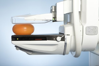
While a growing body of research now suggests that x-ray mammography is causing more harm than good in the millions of women who subject themselves to breast screenings, annually, without knowledge of their true health risks, the primary focus has been on the harms associated with over-diagnosis and over-treatment, and not the radiobiological dangers of the procedure itself.

In 2006, a paper published in the British Journal of Radiobiology, titled "Enhanced biological effectiveness of low energy X-rays and implications for the UK breast screening programme," revealed the type of radiation used in x-ray-based breast screenings is much more carcinogenic than previously believed:

Recent radiobiological studies have provided compelling evidence that the low energy X-rays as used in mammography are approximately four times - but possibly as much as six times - more effective in causing mutational damage than higher energy X-rays. Since current radiation risk estimates are based on the effects of high energy gamma radiation, this implies that the risks of radiation-induced breast cancers for mammography X-rays are underestimated by the same factor.[1]

In other words, the radiation risk model used to determine whether the benefit of breast screenings in asymptomatic women outweighs their harm, underestimates the risk of mammography-induced breast and related cancers by between 4-600%.

The authors continued
Risk estimates for radiation-induced cancer – principally derived from the atomic bomb survivor study (ABSS) – are based on the effects of high energy gamma-rays and thus the implication is that the risks of radiation-induced breast cancer arising from mammography may be higher than that assumed based on standard risks estimates.

This is not the only study to demonstrate mammography X-rays are more carcinogenic than atomic bomb spectrum radiation. There is also an extensive amount of data on the downside of x-ray mammography.

Sadly, even if one uses the outdated radiation risk model (which underestimates the harm done),* the weight of the scientific evidence (as determined by the work of The Cochrane Collaboration) actually shows that breast screenings are in all likelihood not doing any net good in those who undergo them.

In a 2009 Cochrane Database Systematic Review,** also known as the Gøtzsche and Nielsen's Cochrane Review, titled "Screening for breast cancer with mammography," the authors revealed the tenuous statistical justifications for mass breast screenings:

Screening led to 30% overdiagnosis and overtreatment, or an absolute risk increase of 0.5%. This means that for every 2000 women invited for screening throughout 10 years, one will have her life prolonged and 10 healthy women, who would not have been diagnosed if there had not been screening, will be treated unnecessarily. Furthermore, more than 200 women will experience important psychological distress for many months because of false positive findings. It is thus not clear whether screening does more good than harm.[2]

In this review, the basis for estimating unnecessary treatment was the 35% increased risk of surgery among women who underwent screenings. Many of the surgeries, in fact, were the result of women being diagnosed with ductal carcinoma in situ (DCIS), a "cancer" that would not exists as a clinically relevant entity were it not for the fact that it is detectable through x-ray mammography. DCIS, in the vast majority of cases, has no palpable lesion or symptoms, and some experts believe it should be completely reclassified as a non-cancerous condition.

A more recent study published in the British Medical Journal in 2011 titled, "Possible net harms of breast cancer screening: updated modeling of Forrest report," not only confirmed the Gøtzsche and Nielsen's Cochrane Review findings, but found the situation likely worse: 

This analysis supports the claim that the introduction of breast cancer screening might have caused net harm for up to 10 years after the start of screening.[3]

So, let’s assume that these reviews are correct, and at the very least, the screenings are not doing any good, and at worst, causing more harm than good. The salient question, however, is how much more harm than good? If we consider that, according to data from Journal of the National Cancer Institute (2011), a mammogram uses 4 mSv of radiation vs. the .02 mSv of your average chest x-ray (which is 200 times more radiation), and then, we factor in the 4-600% higher genotoxicity/carcinogenicity associated with the specific "low-energy" wavelengths used in mammography, it is highly possible that beyond the epidemic of over-diagnosis and over-treatment, mammograms are planting seeds of radiation-induced cancer within the breasts of millions of women.***

With the advent of non-ionizing radiation based diagnostic technologies, such as thermography, it has become vitally important that patients educate themselves about the alternatives to x-ray mammography that already exist.  Until then, we must use our good sense - and research like this - to inform our decisions, and as far as the unintended adverse effects of radiation go, erring on the side of caution whenever possible.

Additional Reading

Is X-ray Mammography Findings Cancer or Benign Lesions?

The Dark Side of Breast Cancer Awareness Month

Does Chemo & Radiation Actually Make Cancer More Malignant?

*This discrepancy in radiation risk models/estimates follows from two fundamental problems: 1) the older risk model was based on higher-energy radiation emissions, such as are given off from atomic bomb blasts 2) it was a crude model, developed before the discovery of DNA and a full understanding of radiotoxicity/genotoxicity.

** Keep in mind that the Cochrane Database Review is at the top of the "food chain" of truth, in the highly touted "evidence-based model" of conventional medicine. Cochrane Database Reviews are produced by The Cochrane Collaboration, which is internationally recognized as the benchmark for high quality, evidence-based information concerning the effectiveness (or lack thereof) of common health care interventions. The organization, comprised of over 28,000 dedicated people from over 100 countries, prides itself on being an "independent" source of information, and historically has not been afraid to point out the corrupting influence of industry, which increasingly co-opts  the biomedical research and publishing fields.

***The low-energy wavelengths cause double strand breaks within the DNA of susceptible cells, which the cell can not repair. Through time these mutations result in "neoplastic transformation"; radiation has the ability to induce a cancerous phenotype within formerly healthy cells that has cancer stem cell-like (CSC) properties.

[1] Enhanced biological effectiveness of low energy X-rays and implications for the UK breast screening programme. Br J Radiol. 2006 Mar ;79(939):195-200. PMID: 16498030

[2] Screening for breast cancer with mammography. Cochrane Database Syst Rev. 2009(4):CD001877. Epub 2009 Oct 7. PMID: 19821284

[3] Possible net harms of breast cancer screening: updated modelling of Forrest report. BMJ. 2011 ;343:d7627. Epub 2011 Dec 8. PMID: 22155336